Midline or deviated caliber mass. The interpretation of a chest film requires the understanding of basic principles.
 Anatomy Of A Chest X Ray How To Read A Chest X Ray Part 1 Youtube
Technique normal anatomy and common pathology are presented.
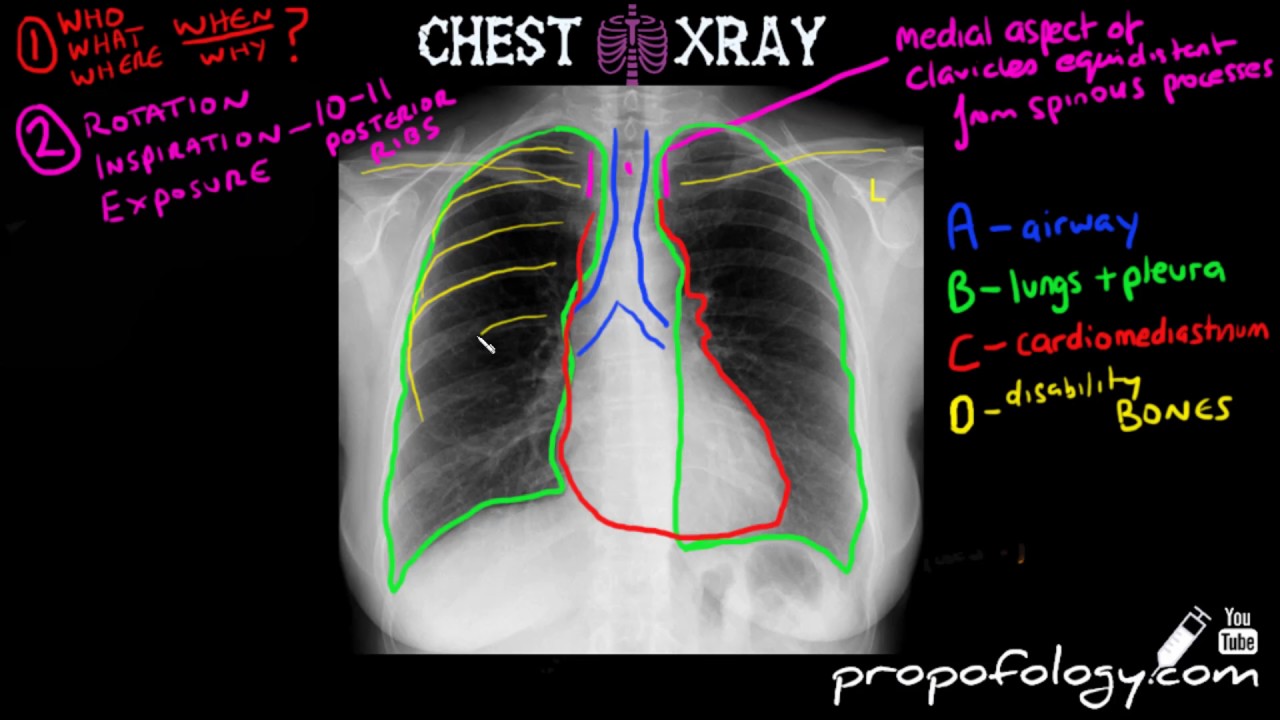
Reading chest x ray. Some may choose to read a chest X-Ray in an anatomical order and some may choose to use a mnemonic. APPA exposure rotation supine or erect. Abnormal shadowing or lucency.
To read a chest x-ray start by looking for markers on it like L for left R for right PA for posteroanterior and AP for anteroposterior to identify the positioning of the x-ray. Ribs are counted from anterior sides. Why order a Chest X-Ray.
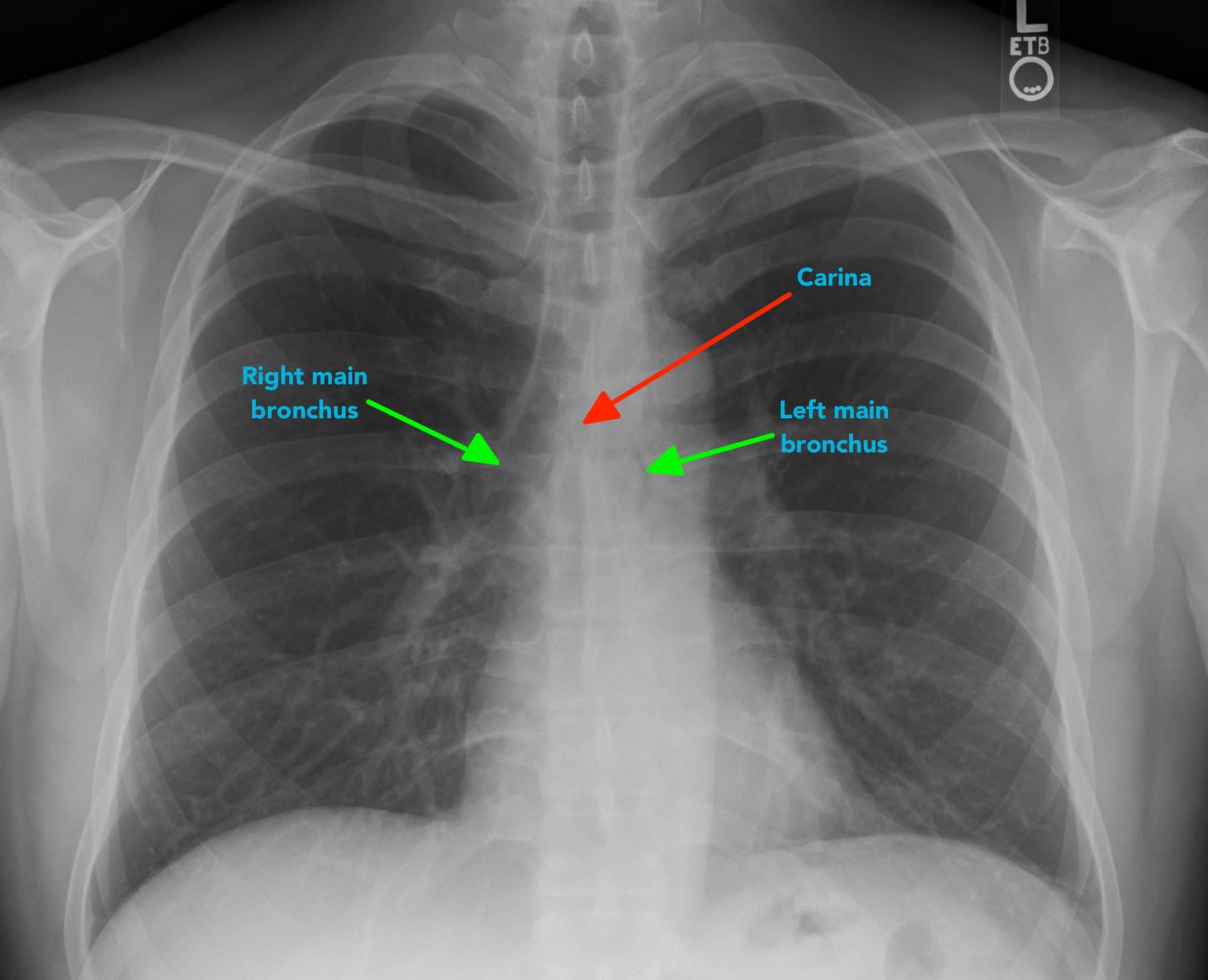
Therefore it is convenient to be able to read and interpret chest x-rays as normal versus abnormal on your own. The chest radiograph is a diagnostic image that is used to see the lungs heart blood vessels airways ribs and spine. Then find the airway on the x-ray and check to see if its patent and midline.
The chest x-ray is the most frequently requested radiologic examination. Systematic approach to the chest film using an inside-out. Inspect the lung zones ensuring that lung markings are present throughout.
If the heart base is 12 the width of the diaphragm on the chest X-Ray it refers to cardiomegaly or pericardial effusion. As a general rule the heart base should not be wider than 12 the total width of the diaphragm. Adult chest x-ray in the exam setting.
Introduction to the Chest X-Ray. This article is an attempt to give the reader guidance how to read a chest Xray and below are two methods. However the important message I would like to give is to adopt one or the other approach and to use the chosen approach consistently.
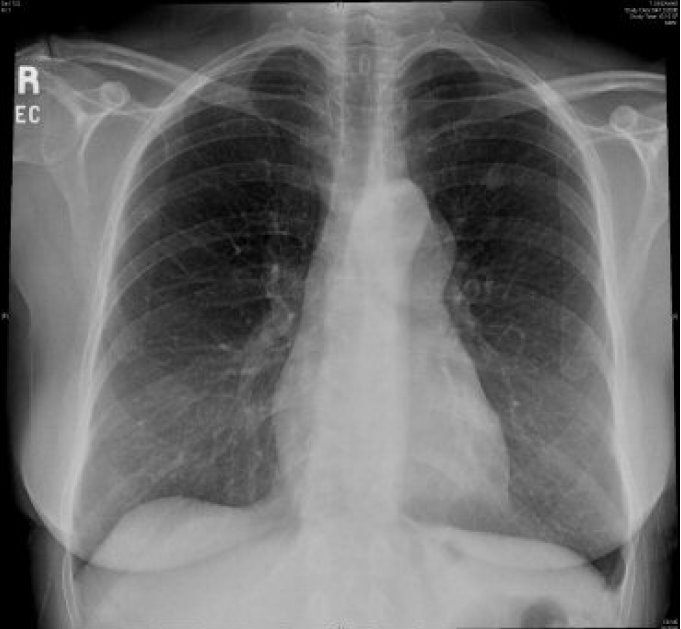
Before we proceed let us see what structures lie in a normal Chest X-Ray. Chest X-Ray Views PA and Lateral. Chest x-ray in the exam setting.
The chest radiograph also known as the chest x-ray or CXR is anecdotally thought to be the most frequently-performed radiological investigation globally although no published data is known to corroborate thisUK government statistical data from the NHS in England and Wales shows that the chest radiograph remains consistently the most frequently requested imaging test by GPs 2019 dataset 5. These zones do not equate to lung lobes eg. Turn off stray lights optimize room lighting view images in order.
Find our complete video library only on Osmosis Prime. The left lung has three zones but only two lobes. There are several reasons why a clinician might order a chest x-ray including trauma cough chest pain or shortness of breath.
But the basics of Chest Xray here will guide you through various aspects including Counting ribs PA vs AP view Inspiratory vs. How to Read a Chest X-Ray. Quizzes are provided for practice and self-assessment.
In this article we will focus on. We have assembled 100 normal Chest X-Rays that were given the Diagnosis of No Active Disease NAD at the Hospital of the University of Pennsylvania HUP. Patients full name and date of.
Reading the chest X-ray systematically reduces the chance of missed diagnosis. The front opaque appearing side of ribs is actually its posterior side. There is no one recommended analysis methodology.
This is an easily overlooked part of the chest x-ray and it should be checked for swelling masses etc. Pediatric chest x-ray in the exam setting. The first in our c.
Patient Data name history age sex old films Routine Technique. Neonatal chest x-ray in the exam setting. This web site is intended as a self-tutorial for residents and medical students to learn to interpret chest radiographs with confidence.
The Chest X-Ray is usually divided into three zones as. By reading this series of Normal CXR students will learn to appreciate the range of normal markings the basics of CXR reading and how patient age and sex influence differentials. When interpreting a chest X-ray you should divide each of the lungs into three zones each occupying one-third of the height of the lung.
Two points can just help you quickly count ribs from top to bottom. Enlargement of the cardiac silhouette. Normal anatomy and variants.
HttposmsitmoreHundreds of thousands of current future clinicians learn by Osmosis. Chest x-rays are common as they are often ordered on patients with chest pain shortness of breath respiratory symptoms concerns for pneumonia etc. This is the cardiac silhouette and theres the atrial appendage the right atrium and the left ventricle.
C is for cardiac silhouette and size. However everyone should begin analysing an X-Ray by checking the following details. It first appears too complicated to read the chest xrays because we barely know what lies where and what to make out of it.
Counting Ribs in Chest X-Ray. The silhouette of the heart should be identified and the heart borders should be clear. While simple learning how to read a chest x-ray is a basic skill all radiologists should know well before moving on to reading more advanced imaging even in the remote reaches of Nepal For patients there are several advantages to using x-rays when possible including lower radiation doses faster acquisition times and lower total cost.
X ray is a type of radiography and most widely used investigation. There is no perfect way to read an x-ray. Basics of Reading Chest X ray.
Reading and interpreting chest x-rays is a useful skill to have no matter what type of specialty you practice. In fact every radiologst should be an expert in chest film reading.
 Learn To Read A Chest Xray In 5 Minutes Youtube
 How To Read Chest X Rays International Emergency Medicine Education Project
 How To Read Chest X Ray Freemedicalmcqs Com
 How To Read Chest X Rays International Emergency Medicine Education Project
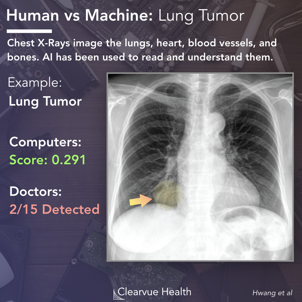 Ai Vs Radiologists Performance On Chest X Rays Visualized Science
 Chest X Ray Interpretation A Structured Approach Radiology Osce
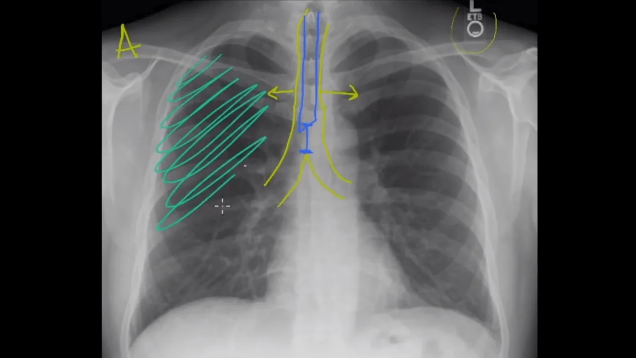
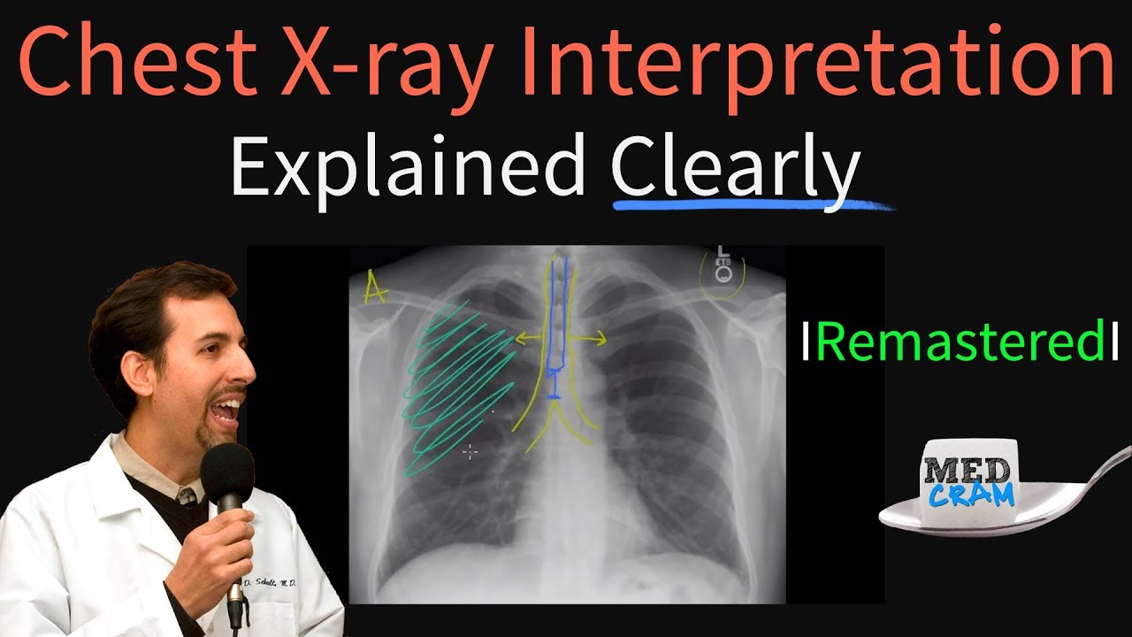 Chest X Ray Interpretation Explained Clearly How To Read A Chest Xray Youtube
 Chest X Ray Interpretation Made Easy Learn To Read A Cxr
 How To Read A Chest Xray Mnemonic Radiology Student Radiographer Medical Education
 The Radiology Assistant Basic Interpretation
Chest X Ray Interpretation A Structured Approach Radiology Osce
![]() Normal Chest X Ray Anatomy Tutorial Kenhub
Normal Chest X Ray Anatomy Tutorial Kenhub
 Reading Chest X Rays Rk Md