101 to 2 mm. Melanoma 20 to 40 mm in thickness ulceration status unknown or unspecified.
 Why Is Melanoma So Dangerous Skin Analytics
Tis melanoma in situ.

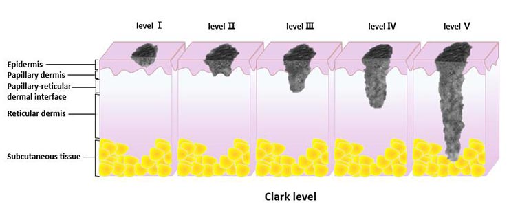
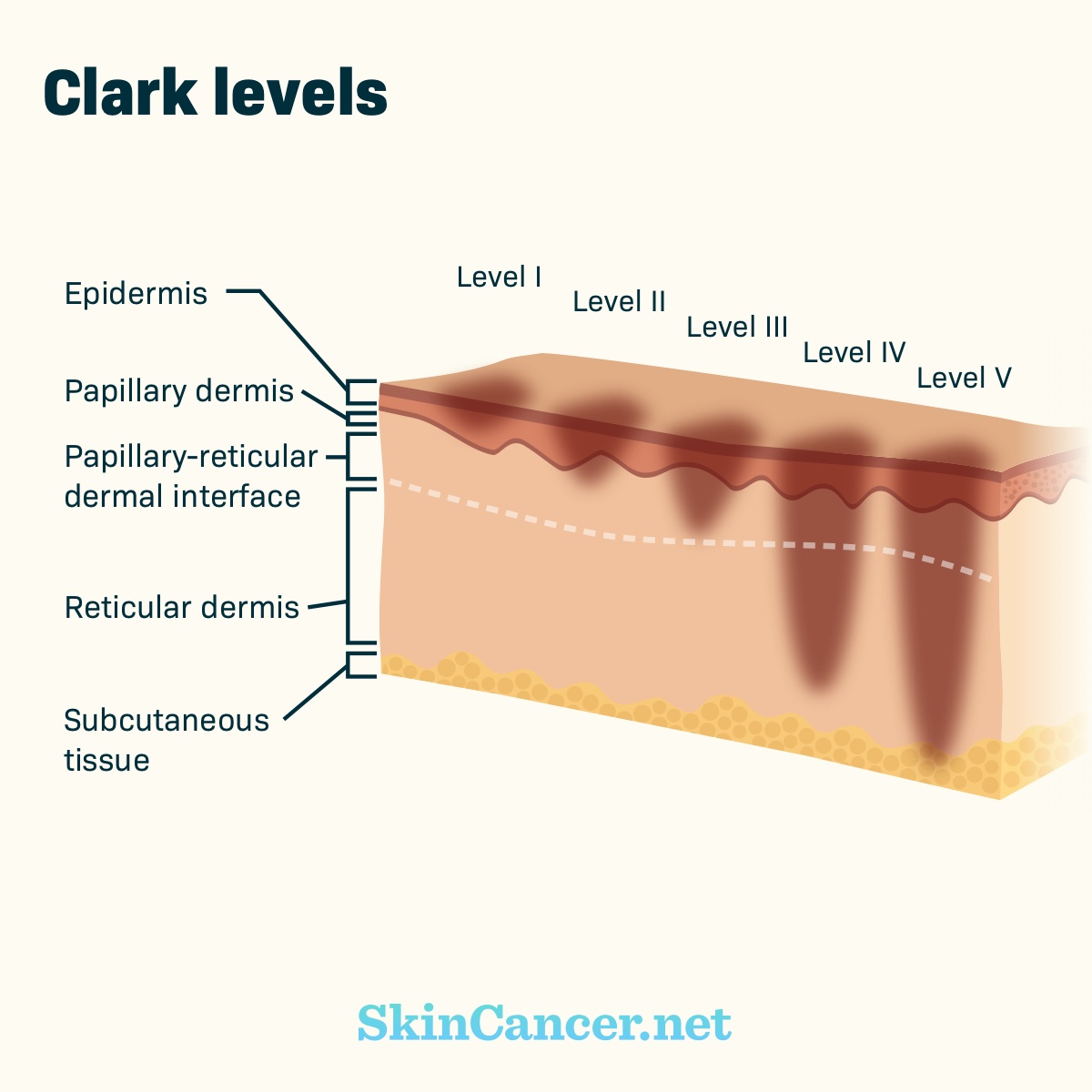
Melanoma clark level 2. Clarks Level also called anatomic level is also a measure of depth of invasion. Melanoma has invaded reticular dermis. Melanoma has invaded subcutaneous tissue.
Clark level I represents intraepidermal or in situ melanoma Fig. Freaking outJust diagnosed on friday with a clark level 2 melanoma on my thigh. Level 1 is also called melanoma in situ the melanoma cells are only in the outer layer of the skin the epidermis Level 2 means there are melanoma cells in the layer directly under the epidermis this is known as the papillary dermis superficial dermis.
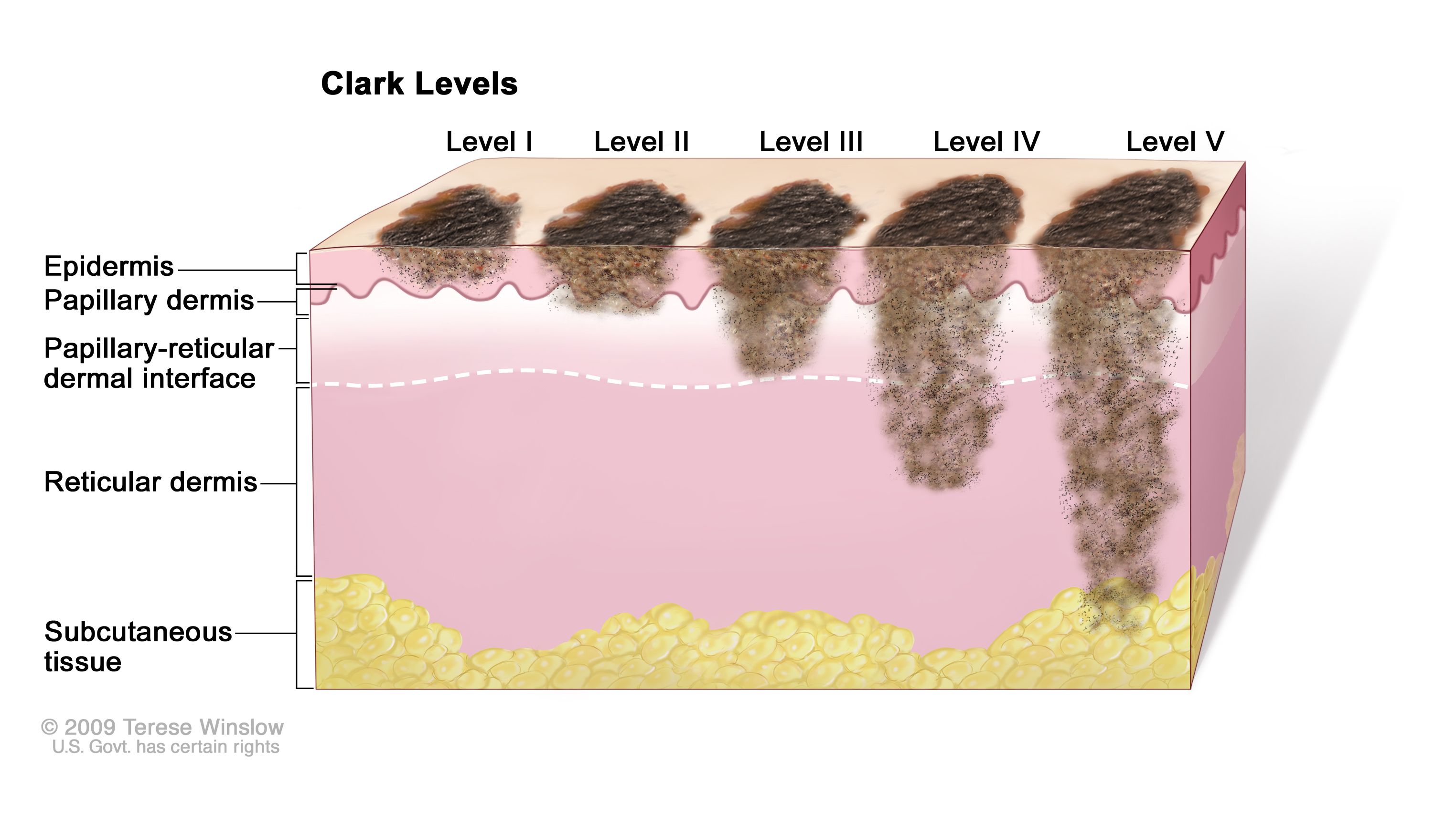
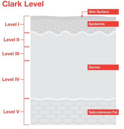
There are five Clark levels of invasion. Clarks Level II means it has moved from the epidermis into the top layer of the dermis nothing more. There is no lymph node involvement or spread to organs.
14 In the most recent version of the AJCC melanoma staging system T1b melanomas were redefined as tumours 1 mm in Breslow thickness with ulceration or a mitotic rate 1mm2 regardless of Clark level. Less than 1 mm. Previously Clark level of invasion was used as an indicator of prognosis but in the AJCC database multivariate analysis showed that it was a much less important prognostic factor.
It may be ulcerated or not ulcerated. There are five Levels within the Clark scale with Level 1 being least developed and Level 5 being most developed. Melanomas confined to the outermost layer of the skin the epidermis.
Clark Level applicable to invasive tumor only Note D. The melanoma cells are only in the top layer of the skin the epidermis. Penetration by melanomas into the second layer of the skin the dermis.
Around 95 meaning 95 people out of 100 people will be alive five years after being diagnosed with a melanoma that is less than 1 mm thick. 201 to 4 mm. The Clark scale has 5 levels.
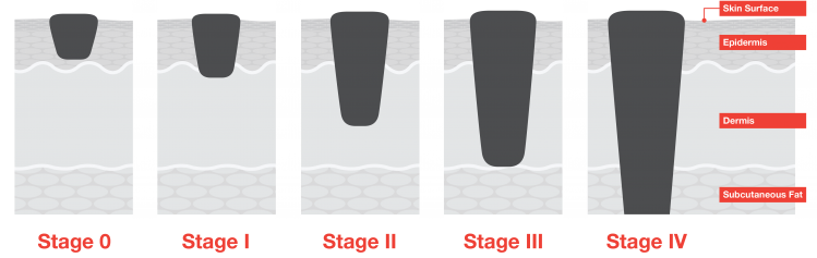
Melanoma is confined to the epidermis the outer layer of the skin. Stage II melanoma is defined by thickness and ulceration. I go in on thursday to have it removed and they said i would just be monitored after removal.
The higher the Clarks Level number the deeper into the tissue it. Your pathology report contains information such as tumor stage Clark level Breslow thickness ulceration occurs when melanoma breaks through the overlying skin and mitotic rate MR. Clarks level is a staging system which describes the level of anatomical invasion of the melanoma in the skin.
Freaking outJust diagnosed on friday with a clark level 2 melanoma on my thigh. It reports what layer of the skin the melanoma extends into penetration of as opposed to a measurement in millimeters. Melanoma has invaded the papillary dermis the outermost layer of the dermis the next layer of skin.
The cells have spread into the layer just beneath the epidermis the papillary dermis. This describes the size of the primary original tumor and whether it has invaded into nearby tissue. Stage 2 melanoma means the tumor is more than 1 mm thick and may be larger or have grown deeper into the skin.
These are the characteristics that determine staging. At Harvard University and Massachusetts General Hospital in the 1960s. Surgical removal is the main treatment however the removal of nearby lymph nodes is also a treatment option to prevent further spread.
Clarks Level II is an outdated method you need to learn the depth of the lesion mitosis ulceration. Clark level III refers to melanomas that fill andor expand the papillary dermis Fig. Melanoma has invaded papillary dermis.
Also called melanoma in-situ Level II. The Clark level indicates the anatomic plane of invasion. After byopsy the depth is a 19 what exactly does this mean stage wise.
Melanoma has filled papillary dermis. The distinction between Clark level 2 and 3 as well as between level 3 and 4 is difficult to apply in practice and lacks reproducibility when compared to Breslow thickness. A high mitotic rate also correlates with a greater likelihood of having a positive sentinel lymph node biopsy.
After byopsy the depth is a 19 what exactly does this mean stage wise. Melanoma has invaded throughout the papillary dermis and is touching on the next deeper layer of the dermis. Clark levels indicate which microanatomic compartments are involved by melanoma.
Melanomas invade deeper through the dermis but are still contained completely within the skin. It has also been found that if the mitotic rate can be assessed in thin melanomas then Clark level does not provide any additional independent prognostic information. The cancer has not spread to nearby lymph nodes.
High risk thick melanoma. Clark level II reflects partial invasion of the papillary dermis Fig. The Clark Scale has five levels.
Greater than 40mm in depth. I go in on thursday to have it removed and they said i would just be monitored after removal. Melanoma In Situ anatomic level 1 ___ Superficial spreading melanoma in situ ___ Melanoma in situ.
It was developed by Wallace H. Greater than 4 mm. When discussing melanoma staging you will see references to the Tumor size Lymph Node involvement and Metastasis.
 Full Text The Relationship Between Mitotic Rate And Depth Of Invasion In Biopsie Ccid
 Definition Of Clark Level Ii Skin Cancer Nci Dictionary Of Cancer Terms National Cancer Institute
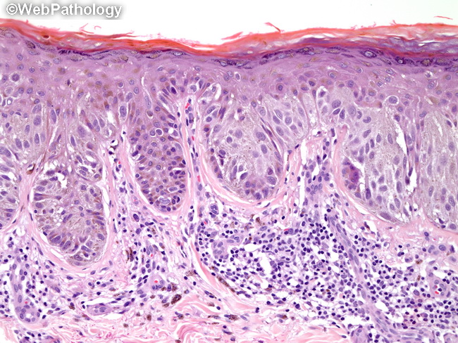 Webpathology Com A Collection Of Surgical Pathology Images
 Clark Level Tnm Stage Download Table
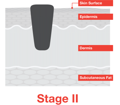 Stage 2 Melanoma Melanoma Research Alliance
 Breslow Depth And Clark Level Melanoma Research Alliance
 Learn To Distinguish Moles From Melanomas Cusabio
/iStock_24463523_LARGE-5809a6205f9b58564cf5ff19.jpg) What Each Melanoma Stage Means
What Each Melanoma Stage Means
Clark Level 4 Melanoma Survival Rate Rating Walls
 Melanoma Staging Melanoma Research Alliance
 Surgicalcore Table Figure

 Clark Level And Breslow Thickness Pharmacotherapy Principles
 Stages Of Melanoma Skincancer Net